PRINCIPLE OF ESR
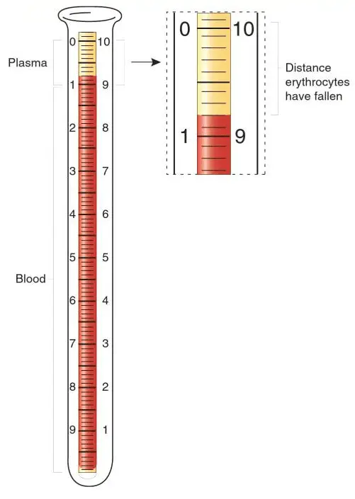
When anticoagulated blood is allowed to stand in a narrow vertical glass tube, undisturbed for a period of time, the RBCs – under the influence of gravity- settle out from the plasma. The rate at which they settle is measured as the number of millimeters of clear plasma present at the top of the column after one hour(mm/hr). This mechanism involves three stages:
- Stage of aggregation : It is the initial stage in which piling up of RBCs takes place. The phenomenon is known as Rouleaux formation. It occurs in the first 10-15 minutes.
- Stage of sedimentation : It is the stage of actual falling of RBCs in which sedimentation occurs at constant rate. This occurs in 30-40 minutes out of 1 hour, depending upon the length of the tube used.
- Stage of packing : This is the final stage and is also known as stationary phase. In this, there is a slower rate of falling during which packing of sedimented RBCs in column occurs due to overcrowding. It occurs in final 10 minutes in 1 hour.
METHOS OF ESR DETERMINATION
There are two main methods to determine ESR :
- Wintrobe’s method
- Westergren’s method
Each method produces slightly different results. Mosely and Bull (1991) concluded that Wintrobe’s method is more sensitive when the ESR is low, whereas, when the ESR is high, the Westergren’s method is preferably an indication of patient’s clinical state.
WINTROBE’S METHOD
This method uses Wintrobe’s tube, a narrow glass tube closed at the lower end only. The Wintrobe’s tube has a length of 11 cm and internal diameter of 2.5 mm. It contains 0.7-1 ml of blood. The lower 10 cm are in cm and mm. The marking is 0 at the top and 10 at the bottom for ESR. This tube can also be used for PCV. The marking is 10 at the top and 0 at the bottom for PCV.
REQUIREMENTS :
- Anticoagulated blood (EDTA, double oxalate)
- Pasteur pipette
- Timer
- Wintrobe’s tube
- Wintrobe’s stand
PROCEDURE :
- Mix the anticoagulated blood thoroughly.
- By using Pasteur pipette, fill the Wintrobe’s tube upto ‘0’ mark. There should be no bubbles in the blood.
- Place the tube vertically in ESR stand and leave undisturbed for 1 hour.
- At the end of 1 hour, read the result.
NORMAL VALUE :
For males : 0-9 mm/hr
For females 0-20 mm/hr
For females 0-20 mm/hr
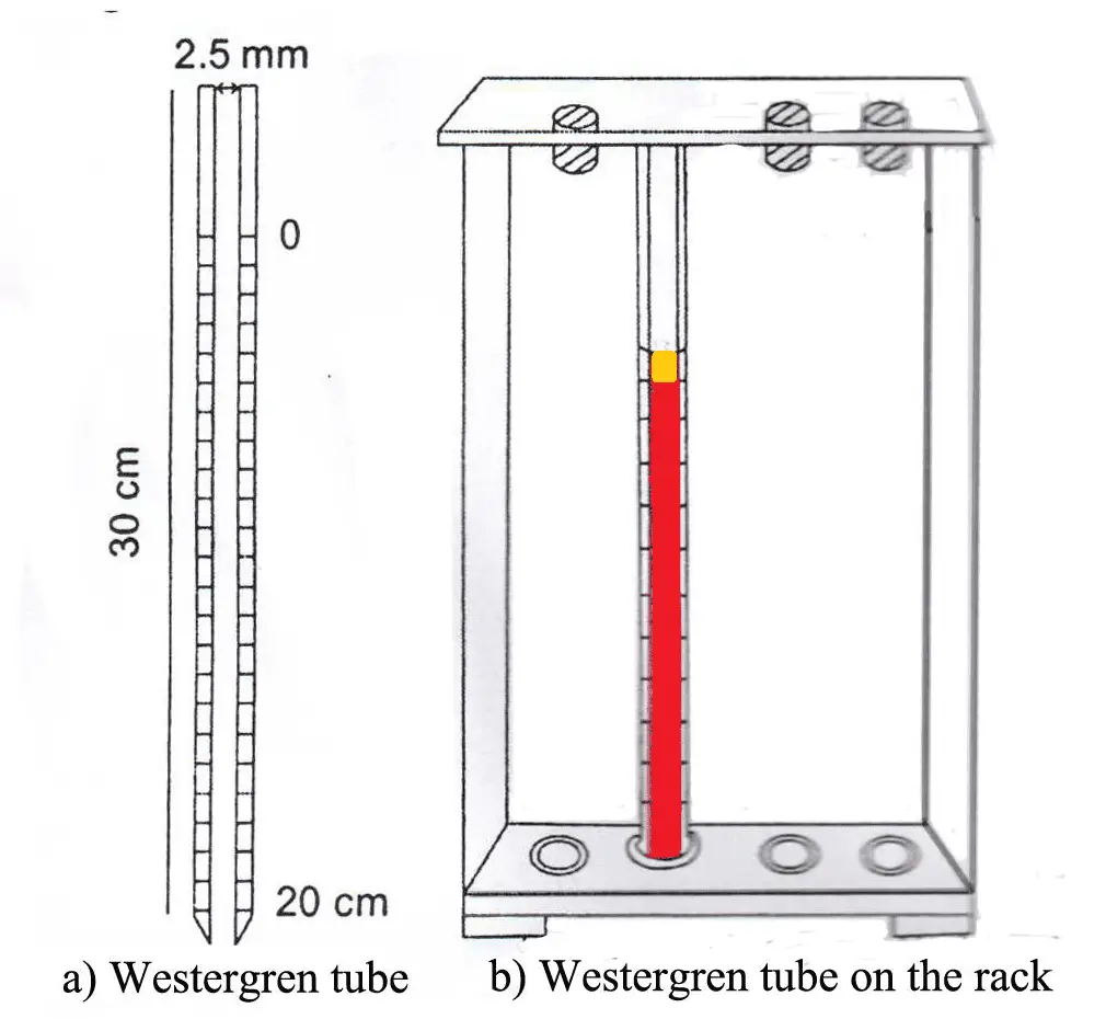
WESTERGREN’S METHOD
It is better method than Wintrobe’s method. The reading obtain is magnified as the column is lengtheir. The Westregren tube is open at both ends. It is 30 cm in length and 2.5 mm in diameter. The lower 20 cm are marked with 0 at the top and 200 at the buttom. It contains about 2 ml of blood.
REQUIREMENTS :
- Anticoagulated blood (0.4 ml of 3.13% trisodium citrate solution + 1.6 ml blood)
- Westergren tube
- Westergren stand
- Rubber bulb (sucker)
PROCEDURE :
- Mix the anticoagulated blood thoroughly.
- Draw the blood into the tube upto 0 mark with the help of rubber bulb.
- Wipe out blood from bottom of the tube with cotton.
- Set the tube upright in stand. Make sure the pipette fits snugly to eliminate possible leakage and that the pipette is in vertical position.
- Leave the tube undisturbed for 1 hour.
- At the end of 1 hour, read the result.
NORMAL VALUE :
For males : 0-10 mm/hr
For females : 0-15 mm/hr
For females : 0-15 mm/hr
Clinical Significance of ESR
The erythrocyte sedimentation rate (ESR) is a non-specific test. It is raised in a wide range of infectious, inflammatory, degenerative, and malignant conditions associated with changes in plasma proteins, particularly increases in fibrinogen, immunoglobulins, and C-reactive protein. The ESR is also affected by many other factors including anaemia, pregnancy, haemoglobinopathies, haemoconcentration and treatment with anti-inflammatory drugs.
Causes of a significantly raised ESR :
- All types of anemias except sickle cell anemia
- Acute and chronic inflammatory conditions and infections including:
– HIV disease
– Tuberculosis
– Acute viral hepatitis
– Arthritis
– Bacterial endocarditis
– Pelvic inflammatory disease
– Ruptured ectopic pregnancy
– Systemic lupus erythematosus - African trypanosomiasis (rises rapidly)
- Visceral leishmaniasis
- Myelomatosis, lymphoma, Hodgkins disease, some tumours
- Drugs, including oral contraceptives
Causes of Reduced ESR :
- Polycythaemia
- Poikilocytosis
- Newborn infants
- Dehydration
- Dengue haemorrhagic fever
- and other conditions associated with haemoconcentration


ليست هناك تعليقات:
إرسال تعليق
Due to the high number of spammy comments we have decided to initiate comment moderation so that we can maintain our quality standards and make good environment for our visitors. Please leave your comment