Principle
Alum acts as mordant and hematoxylin containing alum stains the nucleus light blue. This turns red in presence of acid, as differentiation is achieved by treating the tissue with acid solution. Bluing step converts the initial soluble red color within the nucleus to an insoluble blue color. The counterstaining is done by using eosin which imparts pink color to the cytoplasm.
Reagents
- Harri’s Hematoxylin stain
- Eosin solution
- 0.5% HCl
- Dilute ammonia water
A = 1 gm hematoxylin in 10 ml ethanol
B = 20 gm ammonium alum in hot distilled water
Mix A & B, boil and add 0.5 gm of mercuric oxide and filter.
Yellow eosin = 1 gm
Distilled water = 80 ml
Ethanol = 320 ml
Glacial Acetic Acid = 2 drops
Procedure
- Deparaffinize the section : flame the slide on burner and place in the xylene. Repeat the treatment.
- Hydration : Hydrate the tissue section by passing through decreasing concentration of alcohol baths and water. (100%, 90%, 80%, 70%)
- Stain in hematoxylin for 3-5 minutes
- Wash in running tap water until sections “blue” for 5 minutes or less.
- Differentiate in 1% acid alcohol (1% HCl in 70% alcohol) for 5 minutes.
- Wash in running tap water until the sections are again blue by dipping in an alkaline solution (eg. ammonia water) followed by tap water wash.
- Stain in 1% Eosin Y for 10 minutes
- Wash in tap water for 1-5 minutes
- Dehydrate in increasing concentration of alcohols and clear in xylene
- Mount in mounting media
- Observe under microscope
Result and Interpretation
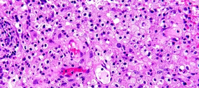
- Nuclei : blue, black
- Cytoplasm : Pink
- Muscle fibres : deep red
- RBCs : orange red
- Fibrin : deep pink


No comments:
Post a Comment
Due to the high number of spammy comments we have decided to initiate comment moderation so that we can maintain our quality standards and make good environment for our visitors. Please leave your comment