Malaria Diagnostic Tests
Malaria Diagnosis (U.S.) - Microscopy
Microscopic examination remains the “gold standard” for laboratory confirmation of malaria. These tests should be performed immediately when ordered by a health-care provider. They should not be saved for the most qualified staff to perform or batched for convenience. In addition, these tests should not be sent out to reference laboratories with results available only days to weeks later. It is vital that health-care providers receive results from these tests within hours in order to appropriately treat their patients infected with malaria.
Technique
(See DPDx specimen preparation) A blood specimen collected from the patient is spread as a thick or thin blood smear, stained with a Romanovsky stain (most often Giemsa), and examined with a 100X oil immersion objective. Visual criteria are used to detect malaria parasites and to differentiate (when possible) the various species. (See DPDx Plasmodium species comparison chart) Wright’s stain, which is commonly used in hospital laboratories for examining blood (called a CBC with manual differential), can be used if Giemsa stain is not available. However, species determination might be more difficult.
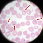
Blood smear from a patient with malaria; microscopic examination shows Plasmodium falciparumparasites (arrows) infecting some of the patient’s red blood cells. (CDC photo)
Advantages
Microscopy is an established, relatively simple technique that is familiar to most laboratorians. Any laboratory that can perform routine hematology tests is equipped to perform a thin and thick malaria smear. Within a few hours of collecting the blood, the microscopy test can provide valuable information. First and foremost it can determine that malaria parasites are present in the patient’s blood. Once the diagnosis is established – usually by detecting parasites in the thick smear – the laboratorian can examine the thin smear to determine the malaria species and the parasitemia, or the percentage of the patient’s red blood cells that are infected with malaria parasites. The thin and thick smears are able to provide all 3 of these vital pieces of information to the doctor to guide the initial treatment decisions that need to be made acutely.
Disadvantages
Microscopy results are only as reliable as the laboratories performing the tests. In the United States, there are, on average, 1700 cases of malaria diagnosed and reported each year. Thus, the average laboratorian does not perform this test regularly, and may not be maintaining optimal proficiency


No comments:
Post a Comment
Due to the high number of spammy comments we have decided to initiate comment moderation so that we can maintain our quality standards and make good environment for our visitors. Please leave your comment