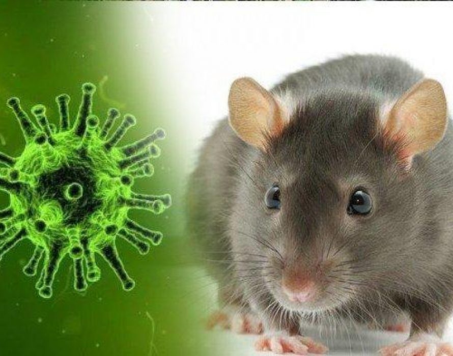
Hantaviruses are rodent-borne viruses causing clinical illness in humans of varying severity. There are several different hantaviruses, with a different geographical distribution and causing different clinical diseases. Each hantavirus is specific to a different rodent host. Transmission of the virus to humans occurs through the inhalation of infected rodent urine, droppings, or saliva.
Three main clinical syndromes can be distinguished after hantavirus infection: haemorrhagic fever with renal syndrome (HFRS), mainly caused by Seoul, Puumala and Dobrava viruses; nephropathia epidemica, a mild form of HFRS caused by Puumala virus; and hantavirus cardiopulmonary syndrome, which may be caused by Andes virus, Sin Nombre virus, and several others. There is no curative treatment for hantavirus infection, and eliminating or minimising contact with rodents is the best way to prevent infection.
1. NAME AND NATURE OF INFECTING ORGANISM
The term hantavirus refers to a genus covering several tens of species or genotypes globally; six so far in Europe, differing in their virulence to humans. Each hantavirus has a specific rodent host species, or a group of closely related host species. Hantaviruses are expanding in Europe: they are found in new areas and the incidence has increased in several established endemic regions.
The most common European hantavirus disease is caused by Puumala hantavirus, carried by the bank vole (Myodes glareolus). The virus is widespread across most of the continent, except for the UK, the Mediterranean coastal regions and the northernmost areas.
Dobrava hantavirus, carried by the yellow-necked mouse (Apodemus flavicollis), is found only in south-east Europe, as far as the Czech Republic and southernmost Germany in the north, though the carrier species has a much wider distribution in Europe to the west and north.
Other hantaviruses in Europe, but with less public health importance, include Saaremaa hantavirus, carried by the striped field mouse (Apodemus agrarius) and found in eastern and central Europe and the Baltic states; Seoul hantavirus, carried by rats (Rattus norvegicus, R. rattus); Tula hantavirus, carried by Microtus voles; and Seewis hantavirus, common in shrews (Sorex araneus), and only recently found in Europe.
Clinical illness results in haemorrhagic fever with renal syndrome (also called “nephropatia epidemica”) and causes less than 0.5% mortality.
YOU MAY ALSO LIKE: Common household cleaning products that are effective against Coronavirus
2. CLINICAL FEATURES
Overall, three syndromes are caused by hantaviruses:
(1) Haemorrhagic fever with renal syndrome (HFRS), mainly in Europe and Asia;
(2) Nephropathia epidemica (NE), a mild form of HFRS, caused by Puumala hantavirus, and occurring in Europe;
(3) Hantavirus cardiopulmonary syndrome (HCPS), in the Americas.
The clinical features in patients with hantavirus disease are quite variable, from asymptomatic to severe. The incubation period is relatively long, mostly 2–3 weeks, but may be up to six weeks. In endemic areas hantavirus infection should be suspected if acute fever is accompanied by thrombocytopenia, headache, often very severe, and abdominal and back pains without clear respiratory tract symptoms.
The case fatality rate due to Puumala virus infection ranges between less than 0.1 and 0.4%. Recovery usually begins during the second week of illness and is accompanied by improvement of urinary output resulting in polyuria. Full recovery may, however, take weeks. Longer-lasting complications are rare, and include glomerulonephritis, Guillain-Barré syndrome, hypopituitarism, and hypertension.
The clinical picture of Dobrava virus infections is very similar, but the symptoms are more severe, with a higher case fatality rate.
3. TRANSMISSION
3.1. Reservoir
Rodents like the bank voles and the yellow-necked mouse are the reservoir for hantaviruses. In the northern part of Europe, human epidemics occur during the cyclic population peaks of the host species. In temperate Europe, on the other hand, human epidemics are related to the (irregular) occurrence of mast years, i.e. years with heavy seed crops of oak and beech leading to abundance of seed-eating rodent species including A. flavicollis. Carrier rodents often invade the human settlements in the autumn thus increasing risk. During rodent peak years, a high proportion of rodents can be seropositive. After being infected, bank voles start to shed the virus after 5–6 days, and the excretion continues for about two months.
3.2. Transmission mode
The rodents excrete hantaviruses in the urine, faeces and saliva, and human infection takes place mostly via inhalation of aerosolised virus-contaminated rodent excreta. Therefore rodent-infested dusty places are risk sites. No human–to-human transmission is known for European hantaviruses. No arthropod vectors are known for hantaviruses.
3.3. Risk groups
Occupations such as forestry workers and farmers have an increased risk of exposure.
4. PREVENTION MEASURES
Avoidance of virus-contaminated dust during work or leisure time is of prime importance; for people with underlying disease, face masks could be used. Creation of air-borne dust should be avoided when areas containing rodent droppings are cleaned, and moist cleaning with disinfectants is recommended. Wild rodents taken into homes as pets or to laboratories for research purposes have caused infections.
Since Puumala virus remains infective outside the host for an unexpectedly long period (for two weeks at room temperature), the risk of infection can persist after rodents have been removed.
YOU MAY ALSO LIKE: How does the novel corona virus attack human cell?
5. DIAGNOSIS
The diagnosis of hantavirus disease mainly relies on the detection of antibodies, through immuno-fluorescent assays (IFA) or Enzyme Immuno Assays (EIA). In the acute phase of the hantavirus infection, antibodies are not specific. Low avidity of IgG antibodies and granular fluorescence in IFA of acute sera can be used to separate old from new infection. In recent years, immuno-chromatographic IgM assays as a point-of-care test with an optical reader, has been developed. RT-PCR from patient blood is coming into use.
6. MANAGEMENT AND TREATMENT
The treatment of hantavirus disease is mainly symptomatic. Maintaining the fluid balance, while avoiding over-hydration in a potentially oliguric patient is of critical importance. In case of renal insufficiency, dialysis may be required. Because European hantaviruses do not spread from human to human, no isolation is needed.
Ribavirin is the only drug used in severe hantavirus infections in Europe. There is currently no vaccine available in Europe.
7. KEY AREAS OF UNCERTAINTY
Hantavirus diseases are under-diagnosed in many regions in Europe; locally adapted guidelines to raise awareness are needed. The respective role of different rodent species in transmitting RBD needs to be further assessed. Rodent vector control strategies need to be further developed and fine-tuned.
YOU MAY ALSO LIKE: Corona virus can remain infective for days in drinking and sewage water
8. REFERENCES
Evander M, Eriksson I, Pettersson L, Juto P, Ahlm C, Olsson GE et al. Puumala hantavirus viremia diagnosed by real-time reverse transcriptase PCR using samples from patients with hemorrhagic fever and renal syndrome. J Clin Microbiol 2007;45:2491–97.
Heyman P, Vaheri A. Situation of hantavirus infections and haemorrhagic fever with renal syndrome in European countries as of December 2006. Eurosurveill 2008;18(28):1–8.
Kallio ERK, Klingström J, Gustafsson E, Manni T, Vaheri A, Henttonen H et al. Prolonged survival of Puumala hantavirus outside the host: evidence for indirect transmission via the environment. J Gen Virol 2006a;87:2127–2134.
Kanerva M, Mustonen J, Vaheri A. Pathogenesis of Puumala and other hantavirus infections. Rev Med Virol 1998;8:67–86.
Klingström J, Heyman P, Escutenaire S, Brus Sjölander K, Dejaegere F, Henttonen H et al. Rodent host specificity of European hantaviruses: characterization of interspecific spillover. J Med Virol 2002;68:581–588.
Lee HW, Chu YK, Woo YD et al. Vaccines against hemorrhagic fever with renal syndrome. In: Saluzzo JF, Dodet B (eds). Factors in the emergence and control of rodent-borne diseases (hantaviral and arenaviral diseases). Amsterdam: Elsevier; 1999, pp. 147–156.
Mustonen J, Partanen J, Kanerva M et al. Genetic susceptibility to severe course of nephropathia epidemica caused by Puumala hantavirus. Kidney Int 1996;49:217–221.
Sauvage F, Langlais M, Pontier D. Predicting the emergence of human hantavirus disease using a combination of viral dynamics and demographic patterns. Epidemiol Infect 2007;135:45–56.
Tersago K, Schreurs A, Linard C, Verhagen R, Van Dongen S, Leirs H. Population, environmental, and community effects on local bank vole (Myodes glareolus) puumala virus infection in an area with low human incidence. Vector Borne Dis 2008b;8:235–44.
Vaheri A, Vapalahti O, Plyusnin A. How to diagnose hantavirus infections and detect them in rodents and insectivores. Rev Med Virol 2008.;18:277–288.
Vapalahti O, Mustonen J, Lundkvist Å, Henttonen H, Plyusnin A, Vaheri A. Hantavirus infections in Europe. Lancet Inf Dis 2003;3:653–661.


ليست هناك تعليقات:
إرسال تعليق
Due to the high number of spammy comments we have decided to initiate comment moderation so that we can maintain our quality standards and make good environment for our visitors. Please leave your comment